
Acute Pericarditis ECG is an important finding to understand because it mimicks STEMI.
Inflammation of the pericardium occurs due to various causes and characteristically presents with chest pain.
ECG in pericarditis is further discussed under following headings:
- Acute Pericarditis
- Chronic pericarditis with Pericardial Effusion
- Cardiac Tamponade
- Constrictive Pericarditis
Acute Pericarditis ECG
Acute pericarditis is caused due to autoimmune, infective or idiopathic reasons, most common being the idiopathic. Most common cause among the idiopathic reason is viral etiology.
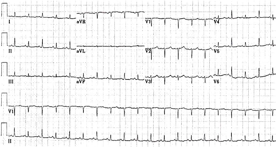
Acute pericarditis ECG findings are:
- ST segment elevation with concave upwards morphology
- TP segment depression with apparent J point elevation due to diastolic injury current
- Taller, Symmetrical and Peaked T-waves
- PTa wave appearance due to atrial injury which is opposite to P wave
- QT interval shortens during acute injury and prolongs during recovry phase
Chronic Pericarditis with Pericadial Effusion ECG
This may be the end result of certain forms of acute pericarditis but may be due to tuberculosis, myxoedema and malignancy.
ECG changes in Chronic Pericarditis
- Diminished amplitude of all the deflections due to fluid acting as a barrier
- T wave inversion due to persistent pericardial injury
- Electrical alternans
- Axis deviations especially Left Axis Deviation
Cardiac Tamponade ECG
Cardiac Tamponade may be a result of acute pericarditis or chronic pericarditis with a large effusion.
ECG changes in Cardiac Tamponade
- Low voltage complexes
- T wave inversion
- Left Axis Deviation
- Electrical alternans is pathognomic of Cardiac Tamponade
- Sinus Tachycardia is invariabl seen in Cardiac Tamponade alongside other arrhythmias
Constrictive Pericarditis ECG
- Constrictive Pericarditis is generally a sequel to pericarditis whether acute or chronic, and is generally characterized by fibrous thickening and loss of elasticity of the pericardium.
- ECG changes in Chronic Constrictive Pericarditis:-
- Low/Inverted/Flattened T waves
- P wave shows Left/Right or Bi-Atrial enlargement
- Low voltage QRS complexes
- The Axis becomes more rightwards with progression of time
- Sinus Tachycardia followed by Atrial Fibrillation are the most common arrhythmias
Pericarditis vs STEMI in ECG
Only non-classical ST elevation in limb leads is not a reliable finding to distinguish acute pricarditis from ST elevation myocardial infarction (STEMI).
- Localised ST elevation can be seen in acute pericarditis but there should be no reciprocal changes in other leads.
- Concave up ST elevation can be found in STEMI.
- But convex up ST elevation is seen only in STEMI.
- ST elevation in lead III which is more than in lead II is strongly suggestive of STEMI.
- PR depression transiently occurs in acute pericarditis and it can also occur in atrial infarction.