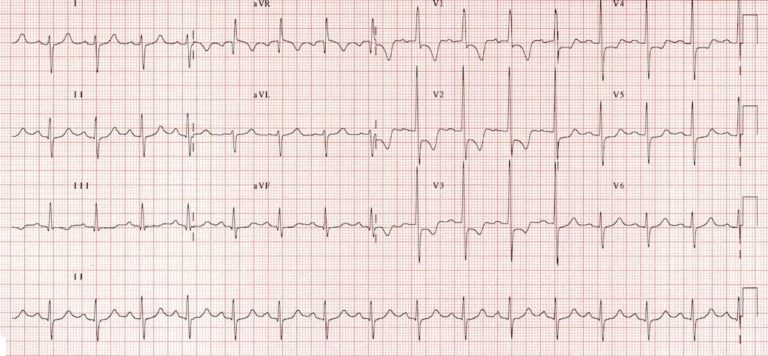
Electrocardiographic Features of Right Ventricular Hypertrophy (RVH)
Right Ventricular Hypertrophy results in the generation of increased QRS forces that are basically directed anteriorly and to the right.
Features of ECG in Right Ventricular Hypertrophy will be covered in this article.
ECG in Right Ventricular Hypertrophy may indicate involvement one or more of three major regions-
- The right paraseptal region
- The free wall of right ventricle
- The basal regions of right ventricle, especially the right ventricular outflow tract

Diagnostic criteria for ECG in Right Ventricular Hypertrophy
- Right axis deviation of +110° or more. degrees
- Dominant R wave in V1 (> 7mm tall or R/S ratio > 1).
- Dominant S wave in V5 or V6 (> 7mm deep or R/S ratio < 1).
- QRS duration <120ms (i.e. changes not due to RBBB).
Supporting criteria for ECG in Right Ventricular Hypertrophy
- Right atrial enlargement (P pulmonale).
- Right ventricular strain pattern = ST depression / T wave inversion in the right precordial (V1-4) and inferior (II, III, aVF) leads.
- S1 S2 S3 pattern = far right axis deviation with dominant S waves in leads I, II and III.
- Deep S waves in the lateral leads (I, aVL, V5-V6).
Other abnormalities caused by RVH
- Right bundle branch block (complete or incomplete).
Note-
There are no universally accepted criteria for diagnosing RVH in the presence of RBBB; the standard voltage criteria do not apply.
However, the presence of incomplete / complete RBBB with a tall R wave in V1, right axis deviation of +110° or more and supporting criteria (such as RV strain pattern or P pulmonale) would be considered suggestive of RVH.
ECG in right ventricular Hypertrophy- Causes of Right Ventricular Hypertrophy
- Pulmonary hypertension
- Mitral stenosis
- Pulmonary embolism
- Chronic lung disease (cor pulmonale)
- Congenital heart disease (e.g. Tetralogy of Fallot, pulmonary stenosis)
- Arrhythmogenic right ventricular cardiomyopathy
Different emphasis of ECG in Right Ventricular hypertrophy in different ventricular regions
1.Dominant hypertrophy of right free wall
- Tall R waves in right precordial leads.
- Mean frontal plane QRS axis deviated inferiorly and to the right: to the region of 120 degrees.
2.Dominant hypertrophy of paraseptal wall
- Tall R waves of RS complexes in the transition zone
- Mean frontal plane QRS axis deviated inferiorly and to the right: to the region of +90 degrees to +120 degrees.
3.Dominant hypertrophy of the right basal regions
- rS complexes in leads V1 to V6 with deep S waves in leads V5 and V6
- qR complex in lead aVR
- Terminal S waves in all three standard leads: the S1, SII, SIII syndrome
- Mean frontal plane QRS axis directed to the right superior quadrant
ECG in Right Ventricular Hypertrophy- Right ventricular hemodynamic overloads
As with left ventricular hypertrophy, the electrocardiographic presentation of right ventricular hypertrophy can vary with the hemodynamic state. The strain on right ventricle may occur during systole or diastole.
1.Right ventricular systolic overload
Right ventricular systolic or pressure overload occurs when there is resistance to right ventricular outflow so thatthe strain on the right ventricle occurs during systolic cintraction. This hemodynamic state is associated with conditions such as pulmonary stenosis and pulmonary hypertension. The electrocardiographic presentation of right ventricular systolic overload is described above, consisting mainly of tall R waves with inverted T waves in the right precordial leads. The U wave may also become diminished in amplitude or even inverted in the right precordial leads as well as in the inferiorly oriented leads: standard leads II and III and aVF.
2.Right ventricular diastolic overload
Right ventricular diastolic overload occurs when there is excessive filling of the right ventricular during diastole. this occurs, for example, with atrial septal defect when there is a moderate to large left-to-right shunt. The electrocardiographic presentation is incomplete or complete right bundle branch block. The rR’ configuration in lead V1 of right ventricular systolic overload is, at times, confused with incomplete right bundle branch block. The diagnosis of incomplete right bundle branch block should only be entertained if there is a distinct rs deflection before the R’ wave, that is an rsR’ complex. The U wave may occasionally become inverted in the right precordialand/or inferiorly oriented leads.
GMCMedicine further readings-
- Junior Resident Doctor Salary in India
- Basic Principles of Arrhythmogenesis
- ECG in Right Ventricular Hypertrophy
- ECG in Coronary Artery Disease
- Next Exam Pattern
External sources to read Right Ventricular Hypertrophy-
