Supraventricular Tachycardia

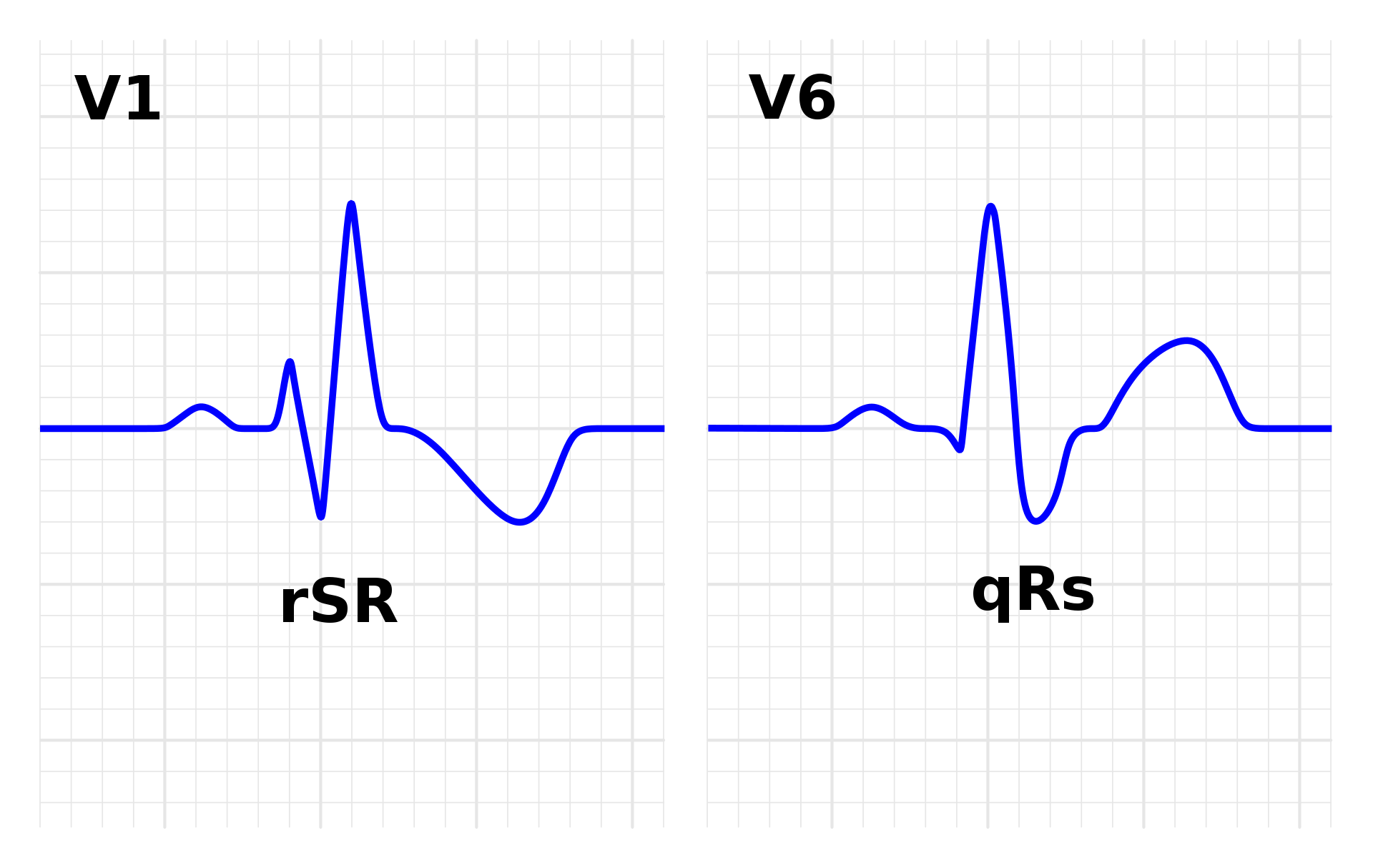
Supraventricular Tachycardia Introduction Tachycardia refers to a heart rate >100 beats per minute (bpm). The tachycardia may be supraventricular or ventricular depending on the origin of the arrhythmia. Supraventricular Tachycardia (SVT): If the tachycardia originates above the bifurcation of the bundle of His, usually in the atria or atrioventricular (AV) junction, the tachycardia is supraventricular. …

Read more

Fascicular Blocks

Fascicular block is a delay or interruption of conduction within one of the major divisions or fasciculi of the left bun­dle branch. The Basic Anatomy for Fascicular Blocks Left Anterior Fascicular Block Common mistakes in left anterior fascicular block: How common left anterior fascicular block is? Symptoms of left anterior fascicular block are? What causes …

Read more

Basic Principles of Arrhythmogenesis

Arrhythmogenesis means genesis of abnormal rhythm of the heart i.e, arrhythmias. Arrhythmias means irregular heart beat. Atrial Fibrillation, Atrial flutter, Some pacemaker requiring abnormal ventricular rhythms are some of the examples of arrhythmias. Basic Principles of Arrhythmogenesis Determination of Heart Rate If RR interval or PP interval is less than three large squares, then the …

Read more

ECG in Right Ventricular Hypertrophy

Electrocardiographic Features of Right Ventricular Hypertrophy (RVH) Right Ventricular Hypertrophy results in the generation of increased QRS forces that are basically directed anteriorly and to the right. Features of ECG in Right Ventricular Hypertrophy will be covered in this article. ECG in Right Ventricular Hypertrophy may indicate involvement one or more of three major regions- …

Read more

ECG in Coronary Artery Disease

Coronary Artery Disease is the most common acquired disease of modern era. Depending on the severity of ischaemia, it can present as- Chronic Stable Angina Chronic Unstable Angina Vasospastic Angina Myocardial Infarction ECG is an important, easily available and reliable tool for diagnosing and planning treatment of a patient with CAD. However, clinical judgement other …

Read more

Basic Principles of ECG

Basic Principles of ECG- P Wave Basic principles of ECG are very important to learn in order to begin reading ECG. No matter how complicated or tough an ECG is, most difficult ECGs can be cracked if one understands the basic principles of ECG. P Wave Overview The P wave is the first positive deflection …

Read more

Sinus Rhythms

Sinus Rhythms may manifest in the following forms:- Normal Sinus Rhythm Sinus arrhythmia Sinus tachycardia Sinus bradycardia Normal Sinus Rhythm Normal sinus rhythm is reflected by the inscription of normal P waves at a rate which ranges between 60 and 100 per minute in the adult. A normal P wave is positive in leads I …

Read more

Left Bundle Branch Block (LBBB)

LBBB ECG :- Left Bundle Branch Block or LBBB is the result of a delay or interruption of conduction within the left bundle branch. In complete LBBB, the duration of QRS is >120ms and in incomplete LBBB it is between 100 and 120 ms. Complete LBBB ECG features :- Prolonged QRS duration >120ms and may …

Read more

Right Bundle Branch Block (RBBB)

Right Bundle Branch Block is a delay or block of conduction within the right bundle branch. In complete RBBB, the duration of QRS is >120ms and in incomplete RBBB it is between 100 and 120ms. Complete RBBB: The ECG Criteria is as follows:- QRS duration >120ms rsR pattern in lead V1 with R’ >r Slurred …

Read more

Wolff-Parkinson-White Syndrome (WPW) – The Great Mimicker

The Wolff-Parkinson-White syndrome is an electrocardiographic syndrome which is the expression of an anomalous atrioventricular conduction pathway, congenital in origin. This pathway forms a bypass which enables the supraventricular impulse to bypass the AV node, bundle of His and distal conducting system, and so activate or pre-excite the ventricles. The anomalous bypass is also known …

Read more

Hypertrophic Cardiomyopathy (HCM)

Hypertrophic Cardiomyopathy is a hereditary disorder that is typically inherited in an autosomal dominant fashion with variable penetrance and expressivity. The hallmark of the disorder is myocardial hypertrophy that is inappropriate, often asymmetrical, and occurs in the absence of an obvious inciting hypertrophy stimulus. This hypertrophy can occur in any region of the left or …

Read more

Cardiac Channelopathies

Ion channels are pore forming proteins that provide pathways for the controlled movement of ions into and out of cells. Diseases caused by mutations in genes that encode ion channel subunits or regulatory proteins are reffered to as channelopathies. Arrhythmogenesis in channelopathies: The heterogeneity in expression of ion channels results in spatial and temporal heterogeneity …

Read more

Electrical Alternans

Electrical Alternans is an electrocardiographic manifestation in which there is alternation in the amplitude of the QRS complex, the T wave alone, OR Both QRS and T wave. It often accompanies fast rates and then has no prognostic significance. When found with slow rates, it connotes organic heart disease with an adverse prognosis. Electrical Alternans …

Read more

ECG in various Drug and Electrolyte Effects

Digitalis Effect ECG in digitalis is manifested as:- Straight downwards slope with a sharp terminal rise like mirror image of a check or correction mark. This does not coonote toxicity and is seen in digitalis therapeutic doses. However, if the beginning of ST segment with the inverse check mark configuration is already depressed below the …

Read more

Emphysema : Chronic Obstructive Airway Disease

The ECG manifestations in a patient of COPD:- Decreaesd magnitude of the electrocardiographic deflections The frontal plane P, QRS and T wave axes are all commonly directed to +90 degree P pulmonale :- best seen in inferior leads and is the expression of right atrial enlargement Right Axis Deviation (RAD) Left Axis Deviation (LAD) may …

Read more

ECG in Acute Pulmonary Embolism

Massive and Acute Pulmonary Embolism results in acute dilatation of the right atrium and ventricle, followed by reflex vasoconstriction of the pulmonary arterioles due to mediators such as serotonin and catecholamines that are released in response to the decreased perfusion of the lungs.

ECG in Mitral Valve Prolapse (MVP)

MVP refers to a systolic billowing of one or both mitral leaflets into left atrium with or without mitral regurgitation. The prevalence of this entity is 1% to 2.5% in the population The disease can be primary or secondary in occurence. The primarry MVP syndrome is autosomal inherited while the secondary causes may be due …

Read more

ECG in Systemic Hypertension

Systemic Hypertension may manifest electrocardiographically in the following manner:- Electrocardiographic signs of left venticular hypertrophy due to systolic overload Electrocardiographic signs of left atrial abnormality Left Axis Deviation because of chronic systemic hypertension ECG features of Left Ventricular Hypertrophy There are deep S waves in the right oriented leads Tall R waves in the left …

Read more

ECG in Heart Failure

Cardiac failure is not a disease but a manifestation of underlying disease of the heart or the circulatory system. Common causes of heart failure are coronary artery disease, cardiomyopathies, valvular heart disease and hypertensive heart disease. A normal ECG is very unlikely in a patient of left ventricular dysfunction and hyas a negative predictive value …

Read more

ECG in Myocarditis

Myocarditis is an under-recognised cardiac disease which can present from asymptomatic form to acute pump failure with gross ECG changes Myocarditis causes a diffuse, but patchy involvement of myocardium including atrium, ventricles and the conducting system. ECG Presentation of Mocarditis:- 1. Abnormalities of QRS complex Increase in QRS duration Bizzare intaventricular conduction defects Pathological Q …

Read more

ECG in Pericarditis

Inflammation of the pericardium occurs due to various causes and characteristically presents with chest pain. ECG in pericarditis is further discussed under following headings :- Acute Pericarditis Chronic pericarditis with Pericardial Effusion Cardiac Tamponade Constrictive Pericarditis Acute Pericarditis Acute pericarditis is caused due to autoimmune, infective or idiopathic reasons, most common being the idiopathic. Most …

Read more

ECG Axis Interpretation

ECG Axis Interpretation Axis interpretation using the schematic illustration demonstrates the relationship between QRS axis and the frontal leads of the ECG. Normal Axis = QRS axis between -30° and +90°.Left Axis Deviation = QRS axis less than -30°.Right Axis Deviation = QRS axis greater than +90°.Extreme Axis Deviation = QRS axis between -90° and …

Read more