Arrhythmogenesis means genesis of abnormal rhythm of the heart i.e, arrhythmias. Arrhythmias means irregular heart beat. Atrial Fibrillation, Atrial flutter, Some pacemaker requiring abnormal ventricular rhythms are some of the examples of arrhythmias.
Basic Principles of Arrhythmogenesis
Determination of Heart Rate
If RR interval or PP interval is less than three large squares, then the rate is over 100 per minute and connotes a tachycardia.
If the RR or PP interval is more than five large squares, then the rate is less than 60 per minute and connotes bradycardia.
Anatomy and Physiology of conducting system
The rate and rhythm of the heart are controlled by the SA Node (Sinoatrial Node) which is situated in the wall of the right atrium to the right of the superior vena caval orifice.
The Sinus impulse once released spreads in the atrial myocardium and is reflected in the ECG as P wave.
Eventually, this impulse reaches the AV Node (Atrioventricular Node), which is situated in the right atrium above the tricuspid valve and just to the right of the interatrial septum.
After a delay at the AV Node, which is reflected in the ECG as the PR interval, it reaches the Bundle of His, then Bundle branches and eventually purkinjee fibre system.
The Bundle of His passes horizontally to the left from the AV node and divides into left and right bundle branches.
Bundle Branches divide into Purkinje fibre system which pierce to the surface of the heart from the Endocardium to the Epicardium.
Pacemakers of the Heart
Virtually every cell of the myocardium, SA and AV Nodes, Bundle of His and every cell of the purkinje system of fibres is capable of acting as a distinct pacemaker. however, SA Node has the maximum intrinsic rhythmicity of 70-80 beats per minute, followed by AV Node and followed by Ventricular Myocardium which has an intrinsic pacemaker activity of 40-60 beats per minute.
Whenever, SA node fails, function of pacemaker is taken up by the AV node.
When SA node and AV node both malfunction, pacemaker function is taken up by the ventricles which is slower and manifests as a ventricular escape rhythm.
Classification of Arrhythmias
Arrhythmias can be classified based on:
- Electrophysiological mechanism of Arrhythmogenesis.
- The anatomic site of origin and pattern of conduction in various cardiac chambers.
Both these classifications of arrhythmias are complementary to each other.
Abnormal Rhythms occur as primary or Secondary disorders. Primary disorders of rhythm reflect a basic, essential abnormality. Secondary disorders of rhythm occur only as a result of, and secondary to, a primary disorder.
Primary Disorders of Rhythm has two categories
- Disturbance of impulse formation
- Disturbance of impulse conduction
Disturbance of Impulse Formation
- Sinus Rhythms:- Sinus arrhythmia, Sinus Tachycardia, Sinus Bradycardia
- Atrial Rhythms:- Atrial extrasystoles, Paroxysmal atrial tachycardia, Atrial flutter, Atrial fibrillation
- Atrioventricular Junctional Rhythms:- AV Junctional Extrasystoles, Junctional ectopic Tachycardia
- Ventricular Rhythms:- VPCs, Idioventricular rhythm, Ventricular tachycardia, Ventricular flutter, Ventricular fibrillation, Reciprocal rhythms.
Disturbances of Impulse Conduction
- SA block
- AV block
- Bundle Branch Blocks and Hemiblocks
- Non-specific intraventricular conduction abnormalities
- The Wolff-Parkinson-White syndrome
Secondary disorders of Rhythm
1. Escape Rhythms
- Atrial Escape
- AV Nodal escape
- Ventricular escape
2. AV dissociation
3. Phasic abberant ventricular conduction
How to describe a cardiac rhythm?
There are three fundamental aspects to every cardiac rhythm:-
- The rhythm has an anatomical origin like SA Node, AV Node or Myocardium or Purkinjee system etc.
- The rhythm has a discharge sequence:- normal inherent discharge, tachycardia, bradycardia, extrasystoles, flutter or fibrillation etc.
- The rhythm has a conduction sequence: for example, 2:1 AV block, complete AV block, 2:1 SA block etc.
Any description of a cardiac rhythm is incomplete without reference to all three of these fundamental aspects.
Basic diagnostic approach to abnormal heart rhythms:
On the basis of the principles mentioned above, cardiac rhythm may be fundamentally analysed as follows:-
- The morphology of P wave is analysed first of all that whether it is normal, ectopic wave, a flutter-F wave, a chaotic fibrillation- f wave.
- Then atrial rate is determined.
- Then regularity of atrial rate is determined.
- The relationship between P wave and QRS complexes is determined.
- The QRS configuration is analysed.
By following th above steps, arrhythmogenesis can be better understoood.
What ChatGPT says about arrhythmogenesis?
Arrhythmogenesis refers to the process or mechanisms that lead to the development and occurrence of cardiac arrhythmias. Cardiac arrhythmias are abnormal heart rhythms, which can be classified into various types, including:
- Tachycardias: These are abnormally fast heart rhythms. Examples include atrial fibrillation (AFib), ventricular tachycardia (VT), and supraventricular tachycardia (SVT).
- Bradycardias: These are abnormally slow heart rhythms. An example is sinus bradycardia.
- Atrial Arrhythmias: These involve irregular electrical activity in the atria of the heart. Atrial fibrillation (AFib) and atrial flutter are common examples.
- Ventricular Arrhythmias: These occur in the ventricles and can be life-threatening. Ventricular tachycardia and ventricular fibrillation fall into this category.
Arrhythmogenesis can occur due to various factors and mechanisms, including:
- Abnormal Electrical Pathways: Disruption or formation of abnormal electrical pathways within the heart can lead to arrhythmias. This can be congenital (present from birth) or acquired due to heart disease.
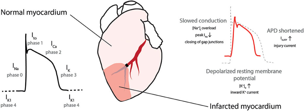
- Ion Channel Abnormalities: Irregularities in the ion channels responsible for regulating the flow of ions (sodium, potassium, calcium, etc.) in and out of heart muscle cells can lead to arrhythmias.
- Heart Disease: Conditions such as coronary artery disease, heart attacks, cardiomyopathy, and heart valve disorders can create a substrate for arrhythmias to develop.
- Medications and Substances: Certain medications, recreational drugs, and even excessive caffeine or alcohol consumption can trigger arrhythmias.
- Electrolyte Imbalances: Disturbances in the levels of electrolytes like potassium, calcium, and magnesium in the blood can affect the electrical activity of the heart and potentially lead to arrhythmias.
- Autonomic Nervous System Dysfunction: Dysregulation of the autonomic nervous system, which controls heart rate and rhythm, can contribute to arrhythmias.
- Genetic Factors: Some arrhythmias have a genetic basis, meaning they can run in families due to specific genetic mutations.
- Age: Aging can lead to changes in the heart’s electrical system, increasing the risk of arrhythmias.
Diagnosis and treatment of arrhythmias typically involve various medical tests, such as electrocardiograms (ECGs or EKGs), Holter monitoring, electrophysiology studies, and imaging studies like echocardiograms. The treatment approach can vary widely depending on the type and severity of the arrhythmia and may include medications, lifestyle changes, electrical cardioversion, catheter ablation, or implantation of devices like pacemakers or implantable cardioverter-defibrillators (ICDs) to regulate heart rhythm and prevent dangerous arrhythmias.


3 thoughts on “Basic Principles of Arrhythmogenesis”