
Sinus Arrhythmias means the rhythms or arrhythmias which originate from the sinoatrial node, travel to atrioventricular node and then via bundle of his and purkinjee fibres activates the ventricles to complete the depolarisation and repolarisation cycle. Because it is following the natural path which is fast and specialised to conduct the impulse in a certain way, this kind of rhythm is referred to as sinus rhythm. If the rhythm is somehow not regular and periodic in time but is originating from sinoatrial node, it is known as sinus arrhythmias.
What is a Normal Sinus Rhythm?
Normal sinus rhythm is reflected by the inscription of normal P waves at a rate which ranges between 60 and 100 per minute in the adult. A normal P wave is positive in leads I and aVL, and in inferior leads, lead aVR is negative and V1 is positive or biphasic with an initial positive component. Normal sinus rhythm is usually associated with normal intraventricular conduction, and is thus reflected by the sequential inscription of P-QRS-T complexes. There is normally an associated slight to moderate sinus arrhythmia.

Sinus Arrhythmias may manifest in the following forms:-
- Sinus tachycardia
- Sinus bradycardia
Sinus Arrhythmias
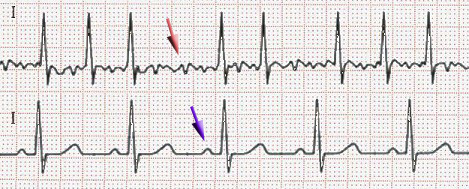
Sinus Arrhythmias are characterized by alternating periods of slow and rapid rates; it is due to an irregular fluctuating discharge of the SA node. The condition is most commonly associated with the phases of respiration – respiratory sinus arrhythmia. The periods of faster rate occur towards the end of inspiration and the periods of slower rate towards the end of expiration. The mechanism is mediated by reflex stimulation of the vagus nerve from receptors in lungs.
Sinus node dysfunction: Sinus node dysfunction can be due to intrinsic or extrinsic causes.
Sinus Arrhythmias- Intrinsic causes of sinus node dysfunction:
Intrinsic disease of the sinus node is associated with structural changes in the sinus node itself or the surrounding atria resulting in progressive deterioration in sinus node function.
This may be due to ischemia, inflammation, infection, infiltrative, metastatic or rheumatic diseases, surgical injury, collagen disease, sclerosis, fibrosis, or idiopathic degenerative diseases that often involve the whole conduction system.
Sinus node dysfunction can be manifested by a number of arrhythmias, although the underlying rhythm disorder is always a bradycardia. About half of all permanent pacemakers in the United States are implanted because of sinus node dysfunction.
Before sinus node dysfunction is attributed to sick sinus syndrome, which is progressive and usually irreversible, extrinsic causes,which are reversible, should be excluded.
Sinus Arrhythmias- Extrinsic and reversible sinus node dysfunction:
The sinus node can be suppressed by neurocardiogenic reflexes; enhanced vagal tone; hypothermia; hypoxia and hypercapnia (especially during sleep apnea); increased intracranial pressure; hypothyroidism; hyperkalemia; and drugs that can suppress the sinus node such as lithium, amitriptyline, clonidine, methyldopa, beta blockers, nondihydropyridine calcium channel blockers, amiodarone, sotalol, and digitalis.
Sinus suppression from extrinsic causes is usually reversible and should be differentiated
from intrinsic disease of the sinus node.
Sinus Arrhythmias Diagnosis:- The PP interval varies alternately with normal P wave morphology and the relationship between P wave and QRS complexes is normal and QRS width is normal.
Significance:- Respiratory sinus arrhythmia is a normal physiological phenomenon and is most marked in young persons. It may cause considerable irregularity of the pulse in childhood.
Some of the specific examples of sinus arrhythmias are:
- Atrial Fibrillation
- Atrial Flutter
- Sinus Bradycardia (Sick Sinus Syndrome)
- Sinus tachy-brady syndrome
- Sinus Tachycardia
- Multifocal Atrial Tachycardia
Sinus Arrhythmias- Sinus Tachycardia
Sinus tachycardia occurs when SA Node discharges at a rate faster than 100 per minute in the adult. The normal resting rate in infants averages 120-130 beats per minute, slowing gradually to reach the adult rate at puberty.
Diagnosis:- The P-QRS-T complexes are recorded in rapid succession. It varies with emotion, respiration and exercise. Vagotonic procedures, for example carotid sinus massage, result in slight and gradual slowing.
Significance:- Sinus tachycardia is the normal physiological response to exercise and emotion. A sinus tachycardia is the normal physiologic response to exercise and emotion. It occurs in anxiety, thyrotoxicosis, toxaemia, cardiac failure and acute carditis. It is a normal accompaniment of fever. The sinus rate will increase by 8 beats per minute for every one degree rise of temperature.
Sinus Arrhythmias- Sinus Bradycardia
Sinus Bradycardia occurs when the SA node discharges at a rate slower than 60 per minute.
Diagnosis:- Sinus bradycardia in the absence of a complicating conduction disturbance, is characterized by normal P-QRS-T complexes, which are recorded in slow succession. It is commonly associated with respiratory sinus arrhythmia.
Significance:- Sinus bradycardia occurs as a normal phenomenon in athletes. Slowing of the sinus rate at times to bradycardiac levels is the physiologic response to sleep. Sinus bradycardia is accentuated by Digitalis and vagotonic procedures, such as carotid sinus compression. The rate quickens gradually with exercise, emotion and amyl nitrate.